![]()
![]()
![]()
![]()
![]()
![]()
![]()
![]()
|
|
|
|
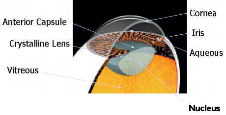
Cataracts & TreatmentWhat is a CataractA cataract is the lens inside the eye that has lost its clarity. As the lens becomes cloudy, vision gets worse. The onset of this is gradual, bit at some point, the loss of vision becomes great enough that an an operation is required to improve the vision. What are the symptoms ?The most common signs of cataracts are foggy or blurred vision. The patient may also notice an increased sensitivity to light and/or a halo effect around lights at night which can make it difficult to drive, caused by light being diffused by the clouding of the lens. Many people have trouble reading, especially small print, colours often appear less bright. TreatmentThe treatment is a cataract extraction. In this procedure, the cloudy lens is removed from the eye. In most situations, an artificial lens, called a lens implant, is placed inside the eye to take the place of the natural lens that has been removed. To understand the different types of cataract operations, it is necessary to know about the structure of the lens. The crystalline lens inside the eye has several parts. The center of the lens is a hard core called the nucleus.
The
lens nucleus is made up of rings of cells, pushed together over time, similar to
the rings in the trunk of a tree. The nucleus starts out very soft in childhood,
but gets progressively harder with age. Surrounding the nucleus is the lens
cortex. The cortex is a softer material. The entire lens is enclosed by the
capsule that is similar to a clear bag. The lens is held in place by small
fibers call the zonule. Several
different procedures have been developed to treat cataracts.
Phacoemulsification - preferred technique for cataract removal Intracapsular Extraction - traditional technique Extracapsular Extraction - traditional technique
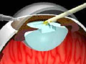
Phacoemulsification
("phaco")
This is now usually the preferred technique for cataract removal.
One of the problems with traditional cataract extraction techniques is that the size of the incision requires significant stitching of the eye, which can lead to scarring and in in some cases astigmatism. Phacoemulsification ("phaco") was developed in the search for a way to extract cataracts through a smaller incision.
It has become the preferred
technique for cataract extraction. A 3mm incision is all that is required. An ultrasound or laser probe is used to break
the lens apart without harming the capsule. These fragments are then sucked (aspirated)
out of the eye. A foldable intraocular lens (IOL) is then introduced through the
3mm incision. Once inside the eye, the lens unfolds to take position inside the
capsule. No stitches are required, as the incision is self-sealing. The risk of
astigmatism and sudden pressure changes inside the eye are minimized. The
procedure is safe enough to be done using anesthetic eyedrops. No injections are
required. Visual rehabilitation is extremely fast and patients don't need to
suspend their everyday activities.
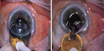
Stages in Phacoemulsification Procedure
A very small cut is made in the outer skin of the eye, (the sclera) using needle-like probe.
A small ultra sound probe is introduced into the cataract and used to completely liquefy it (a laser is sometimes used instead of ultra sound).
A hollow needle is used to suck the liquefied cataract out of the eye, leaving the lens capsule intact.
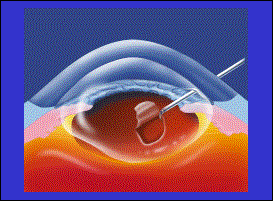
Intracapsular
Extraction
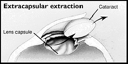
In an intracapsular cataract operation, the entire lens is removed with its capsule.
Intracapsular
cataract operations are now relatively rare. They were the most common type of cataract
surgery until the early 1980s.
Extracapsular Extraction
Some Developments in Cataract Surgery
Topical
Anesthesia (No Injection, and No Patch)
One of the latest and most significant developments in
cataract surgery has been in the use of Topical Anesthesia. In the past
many people would say that the worst part of their cataract operation was
getting an injection "in their eye" to numb it up. Now the entire
surgery (which lasts only about 15 minutes) can be done without an injection,
using only eye drops. The advantages of Topical Anesthesia are numerous. Most
importantly, the patient is spared the pain and risks involved with the injection, plus
the operative results are much faster, and sometimes immediate. Some patients can see better as soon as the
operating microscope is removed. After Topical Anesthesia, the patient can go home without an eye
patch. Also, when combined with a clear corneal incision the patient does not have to stop
taking other medication ( such as aspirin). The use of topical anesthesia eliminates the need for an injection, and it also allows patients to leave the operating room without even a patch.
Temporal
Clear-Corneal Incision (No Stitch)
Even the most modern techniques in cataract surgery require
that an incision be made in the eye. Cataract surgery cannot be performed with
lasers (a common misconception; so called secondary cataracts can be treated
with laser, but secondary cataracts only occur in eyes that have already had
cataract surgery). There appears to be no substantial disadvantage to the temporal, clear-corneal incision.
Foldable
Lens Implant
The final step in cataract surgery is lens
implantation. Prior to the development of safe intra-ocular lens implants,
anyone who had their cataracts removed was forced to wear incredibly thick and
heavy glasses, or contact lenses to correct their vision to normal. A typical three
piece lens implant looks like a miniature, round magnifying glass, about 6
millimeters in diameter, with two wiry attachments called haptics. The haptics
extend out to a total diameter of about 13 millimeters, and when slightly
compressed, they suspend the lens implant inside the lens capsule which itsef has a diameter of
12 millimeter. Obviously this will not fit through
a 3 millimeter incision. Flexible, or foldable lens implants however, can be rolled up into special insertion devices and "injected" through tiny 3 millimeter micro incisions. Once unfolded inside the eye, the haptics suspend the implant inside the same space formerly occupied by the cataract. The haptics heal into place after a few weeks,and further stabilize the implant. Before cataract surgery, the
surgeon makes measurements on
the eyes that assist him or her in selecting the correct lens power. Usually the
power is selected to optimize your distance vision. In other words, by
controlling the power of your implant, your surgeon can correct any pre-existing
near-sightedness or far-sightedness.
New implants are now available that can correct astigmatism, or that can simultaneously correct both distance and near vision. |
|
[Legal] [ Privacy Statement] [Home] [Useful Links ] [Contact Us] Send mail to
Web Team with questions or
comments about this web site.
|